Podcasts


Eye Vitamins 2 [Show Notes]

Remember: if a vitamin is good for one place in your body, it’s probably good for a lot of places in your body.  It’s a marketing ploy to call them eye vitamins.

Eye Vitamins

  1. Thiamine (B-1): thiamine is used heavily in your liver, to help metabolize alcohol.  Most well-known deficiency in alcoholics.  Thiamine also helps nerves produce several neurotransmitters, as well as protects your nerves from inflammatory chemicals (the chemicals in your body that trigger swelling and pain).  You don’t want inflammation in your optical nerves.  You also don’t want the signals from your eyes to your brain and back to be slow.
  2. Folic acid:  important for pregnancy women and fetal development, deficiency can cause a type of anemia.  It’s main job is to help make accurate copies of the DNA and RNA when cells are dividing and multiplying.  You don’t want typos in your DNA!  This is important for your eyes because the cells of your eyes is because they are some of the fastest reproducing and dividing cells in your body.
  3. Omega-3 Fatty acids:  Beneficial for heart health, found in oils.  Oils in your body are lubricating.  Omega-3 fatty acids are building blocks for the cholesterol that is build into cell membranes that keep the fluid and slippery. The body also uses O3FA to produce the natural lubrication in your eye.
  4. N-Acetylcystine (NAC): an amino acid (building block of protein).  Glutathione – your body’s naturally produced antioxidant.  NAC is used when the cells build and store glutathione.  Glutathione focuses mainly on oxygen-based free radicals.
  5. Alpha Lipoid Acid (ALA): another antioxidant.  Found in sources of natural oils – seeds and nuts.
  6. Lutein and Zeaxanthin: 2 nutrients that are found in red, orange, and yellow fruits and vegetables (and that actually cause them to be those colors in the first place).  Their main goal in your eyes are to prevent blue light damage.  Blue light is a short wavelength with a higher speed and energy than the lower colors of light.  This energetic light can damage cells in your eyes, so the orange nutrients blocks this energetic waves so you can still see the blue colors, but it’s more chill and doesn’t cause damage to the retina.  These nutrients concentrate in the macula.

Connect with me

Support us on Patreon

*NEW* Join the Pharmacist Answers Podcast Community on Facebook

Subscribe: iTunesStitcherGooglePlayTuneIn Radio

Like the Facebook page

Music Credits:  “Radio Martini” Kevin MacLeod (incompetech.com)  Licensed under Creative Commons: By Attribution 3.0  http://creativecommons.org/licenses/by/3.0/


Eye Vitamins 1 [Show Notes]

*Disclaimer: most vitamins and minerals are good for ALL of your body.

Eye Vitamins

  1. Vitamin C: helps make collagen, allows iron to be absorbed, and acts as a neurotransmitter co-factor (helps in the process of creating and sending messages).
  2. Vitamin E: antioxidant, it traps up free radicals so they don’t damage cells in important organs.
  3. Beta-Carotene: a pre-cursor to Vitamin A (this happens in your liver). Vitamin A works with proteins in your eyes to create light-sensitive molecules to aid in color vision and seeing in dim light.
  4. Zinc: helps Vitamin A know where it’s needed in the body and helps it get there.
  5. Selenium: helps the body absorb Vitamin E.
  6. Calcium: vital for muscle and nerve conduction (think electricity).
No lone rangers here!
Many foods are fortified in modern countries and have vitamins added to them that may not be naturally occurring in the raw ingredients.  If you’re eating a well balanced diet and still deficient in something, take a supplement of the thing you’re deficient in, not a whole multi-vitamin.  If you’re getting regular check-ups with your doctor, they should be testing for many things, including many vitamin levels, to check for deficiencies.
Being “tired” isn’t always fixed by taking vitamins.

Bonus

Depending on what nutrient is missing to cause anemia, the red blood cells will have a certain appearance.

Some vitamins are fat soluble. They hang out in your adipose tissue, and can cause problems if you get them in too large amounts.

Connect with me

Support us on Patreon

*NEW* Join the Pharmacist Answers Podcast Community on Facebook

Subscribe: iTunesStitcherGooglePlayTuneIn Radio

Like the Facebook page

Music Credits:  “Radio Martini” Kevin MacLeod (incompetech.com)  Licensed under Creative Commons: By Attribution 3.0  http://creativecommons.org/licenses/by/3.0/

Eye Anatomy [Show Notes]

Eyes

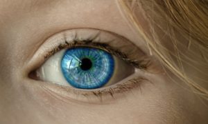
Your eyes are more complex than any camera on the planet!

Cornea:  a concave lens on the front of your eye that focuses light
Iris:  the colored part, a diaphragm that controls how much light comes in (the pupil is the hole the light enters = equivalent to the aperture of a camera)
Lens:  the “focuser”, uses a process called accommodation to focus near to far and make the image as sharp and clear as possible
Retina:  the sensor, and sends signals to the brain to translate light into an images

The retina has 2 types of sensors:

  • Rods – detect light intensity
  • Cones – color differentiation

Two special areas of the retina:

  • Macula – right in the middle of the retina, they place that detects the most detail (that’s why the center of your vision field is a clearer picture than the periphery)
  • Fovea – the center of the macula, it contains cones (color sensors) only to aid in the translation of very fine details

Support structures

  • Extra-ocular muscles – allows your eyes to move around in their holes
  • There are chambers of fluid that are between each structure of the eye, and that fluid helps hold nutrients that feed those parts, and remove waste
  • Choroid:  the layer that holds all the blood vessels that feed the eyes
  • Sclera:  the whites of your eyes, an outer coating that hold everything inside
  • Conjuntiva:  the mucus membrane that attaches the sclera to the eyelids; produces liquid for lubrication and trapping invaders

PSA

Please don’t vigorously rub or scratch your eyes, you could hurt them!

Connect with me

Support us on Patreon

*NEW* Join the Pharmacist Answers Podcast Community on Facebook

Subscribe: iTunesStitcherGooglePlayTuneIn Radio

Like the Facebook page

Music Credits:  “Radio Martini” Kevin MacLeod (incompetech.com)  Licensed under Creative Commons: By Attribution 3.0  http://creativecommons.org/licenses/by/3.0/


Brainstem

An out of focus image of white and colored dots of light. The picture was taken this way, don't blame the out of focus on your brainstem.

Brainstem Basics

Your brainstem is the most basic area of the brain.  The area of the brain that we have in common with almost all other levels of the animal kingdom.  It extends right into the spinal cord.  A lot of other whole body involuntary reflexes come from the spinal cord – that’s another story for another day).

3 main parts

  • Medulla oblongata – rhythm center (heart rate, breathing, swallowing, vomiting and coughing reflex) – they’re all involuntary
  • Pons (not ponds) – the bridge between the cerebellum hemispheres and other brain regions, helps coordinate the right side and left side of your body for complex activities
  • Midbrain – sensory reflexes (also involuntary) – blinking, eye focusing, pupil dilation in response to light, visual and auditory startling reflex that kick-starts the “fight or flight cascade”.

Other eye focusing problems are not rooted in the midbrain. They are more likely rooted in the areas of the brain that control orbital muscles or in the areas that translate what your eyes are seeing, like a “lazy eye” or drifting eye or being cross-eyed.  There are therapies that doctors prescribe to try and strengthen the weak eye.  Blinking is usually a response to eye moisture.

Connect with me

Support us on Patreon

*NEW* Join the Pharmacist Answers Podcast Community on Facebook

Subscribe: iTunesStitcherGooglePlayTuneIn Radio

Like the Facebook page

Music Credits:  “Radio Martini” Kevin MacLeod (incompetech.com)  Licensed under Creative Commons: By Attribution 3.0  http://creativecommons.org/licenses/by/3.0/

Blood Brain Barrier [Show Notes]

Basics

The blood brain barrier is the last layer of cells between what’s in your blood and the extracellular fluid around your brain cells.

You’re born with it!  It’s main job is filtration.

2 ways things get through the blood brain barrier:

  • Passive diffusion: small, neutral molecules (water, gases, lipid-soluble)
  • Active transport: glucose, amino acids, drugs (like a revolving door)

Permeability: how easily something can pass through a layer without work

Things that change permeability:

  • Inflammation – stretches layer and makes holes bigger (meningitis, injury)
  • Multiple sclerosis – an auto-immune disease that can degrade the BBB
  • Alzheimer’s – BBB becomes overwhelmed with antibodies and burns out

*Scary Section*

Rabies is a virus that is small and can get through the blood brain barrier but the immune system cells, antibodies from the vaccine, and medicines can’t.
HIV encephalopathy is caused when a mutation of the HIV gets into the brain and use brain cells to replicate (rather than the well-known T-cells of the immune system).  There is also a rare symptom of HIV called HIV-associated dementia.

Callback

microchimerisms – Pregnancy causes the permeability of many areas of the body to change, and this includes the BBB.

Test Yourself

Drugs that have central nervous system effects (good) or side effects (bad) cross the BBB.  See what you know of different medications and what job they’re supposed to do and what negative side effects they cause and see if you can guess if they cross the BBB.

Connect with me

Support us on Patreon

*NEW* Join the Pharmacist Answers Podcast Community on Facebook

Subscribe: iTunesStitcherGooglePlayTuneIn Radio

Like the Facebook page

Music Credits:  “Radio Martini” Kevin MacLeod (incompetech.com)  Licensed under Creative Commons: By Attribution 3.0  http://creativecommons.org/licenses/by/3.0/


Male DNA in the Female Brain

*Sorry again for crying baby*

Male DNA In The News

The news reported that scientists had discovered a link between male DNA found in the brain of the women who gave birth to sons.

Microchimerism = DNA fragment of another organism that incorporates into you

This particular microchimerism involves the Y chromosome (because otherwise, you wouldn’t know it was specifically male).

Other documented microchimerism studies have reason to believe they may be beneficial – especially in a process called immuno-surveillance (when the immune system is patrolling around looking for things that don’t belong there).

The blood brain barrier is the last layer of cells between what’s in your blood and your brain cells.  DNA fragments are small and can easily pass through the BBB, especially during pregnancy when membrane permeability (the penetrable-ness) has increased all throughout the body already.

The primary resource written by the scientists that did the study of the female brains states that their findings were pretty much inconclusive – partly due to the small sample size of brains they had available.  And they couldn’t study living people.  They were mostly trying to decide if this male microchimerism had a positive or negative effect on Alzheimer’s risk.  The final conclusion – we dunno.  Another obstacle was that the complete health history of the samples they used was not known.

Other sources have stated hypotheses regarding the number of children a woman has and the risk of early onset Alzheimer’s.

This issue with reporting on studies like these is that Alzheimer’s has so many factors that may increase or decrease risk and science is pretty sure there’s NOT just one thing that will cause or prevent someone from developing this disease.

The only thing they could conclude is that microchimerisms are evolutionarily significant.

Here’s the primary journal article.

Connect with me

Support us on Patreon

*NEW* Join the Pharmacist Answers Podcast Community on Facebook

Subscribe: iTunesStitcherGooglePlayTuneIn Radio

Like the Facebook page

Music Credits:  “Radio Martini” Kevin MacLeod (incompetech.com)  Licensed under Creative Commons: By Attribution 3.0  http://creativecommons.org/licenses/by/3.0/


An Accordion in Your Brain

Cerebellum Basics

Your cerebellum is a separate part of your brain that sits under the occipital lobe.  It is responsible for unconscious motor functions, and is organized differently than the cerebrum.  It is packed tightly together in neat folds like an accordion.  And it has 3 lobes:

  • Anterior (in the front) – it keeps the body visually “centered” and on balance, as well as moving the head or body to keep the eyes level with the horizon.  Alcoholism can cause damage to this area that results in a person being sober but still walking “drunk”.
  • Follcular-nodular (in the middle with nodules on it) – responsible for eye movement in response to motion.  It is responsible for correcting balance based on signals from the body rather than the eyes. This is how you know you’re falling over when you have your eyes closed.  Also responsible for muscle tone (aka the passive contraction or “readiness” of a relaxed muscle).
  • Posterior (in the back) – responsible for fine motor coordination and it turns off signals for involuntary movements.

Purkinje cells are the main type of neuron in the cerebellum – SO BEAUTIFUL!!

Connect with me

Support us on Patreon

*NEW* Join the Pharmacist Answers Podcast Community on Facebook

Subscribe: iTunesStitcherGooglePlayTuneIn Radio

Like the Facebook page

Music Credits:  “Radio Martini” Kevin MacLeod (incompetech.com)  Licensed under Creative Commons: By Attribution 3.0  http://creativecommons.org/licenses/by/3.0/


Now You See It [Show Notes]

Occipital Lobe Review

A brain lesion is a place in the brain that doesn’t fire when it should or fires sporadically when it shouldn’t.  Occipital lobe lesions can lead to hallucinations that range from amorphous to extremely detailed.

Field blindness: a lesion causes the occipital lobe to not translate the information from one or more spots of the visual map (your whole view).  Blind spots (round) or visual cuts (lines).

Photosensitivity seizures: seizures triggered by visual overstimulation.  Even though stereotypical in different forms of entertainment, these only accounts for about 10% of seizure triggers.  Seizures triggered by visual stimulation can range from mild to severe.

Certain types of blindness can be rooted in translation problems in the brain, rather than reception problems in the eyeball.

Lesions in the occipital-temporal-parietal junction:

  • Color agnosia: can see the colors but can’t recall the names; simplified colors (all greens appear to be the same green)
  • Movement agnosia: think weeping angels (things only move to a new position when you’re not looking at it) or moving items appear blurry
  • Agraphia: unable to communicate in writin

Connect with me

Support us on Patreon

*NEW* Join the Pharmacist Answers Podcast Community on Facebook

Subscribe: iTunesStitcherGooglePlayTuneIn Radio

Like the Facebook page

Music Credits:  “Radio Martini” Kevin MacLeod (incompetech.com)  Licensed under Creative Commons: By Attribution 3.0  http://creativecommons.org/licenses/by/3.0/

Eyes In The Back of Your Head [Show Notes]

Occipital Lobe Basics

The occipital lobe sits in the back of your head, it directly connects to your eyes.

2 Streams of messages that your eyes send to your occipital lobe.

  1. Ventral stream – translates “what”
  2. Dorsal stream – translates “where” and “how”

It sends translated information to the necessary part of the brain to respond or react to what you saw.  This is how hand-eye coordination works (not just for athletes).  And, since so much of the information we receive is visual means that the occipital lobe doesn’t do much else.

**The following is complete speculation based on my experiences as a Mom.**

Mom’s get accused of having eyes in the back of their head – but my guess is that mom’s gain a keener sense of spacial awareness regarding the things that are happening around you.  Also, mom’s hearing become much more attuned to specific sounds (aka knowing their baby’s voice from other baby voices) to the point of knowing the difference between the sound of crayons coloring on paper versus crayons coloring on a wall!

If it hasn’t been obvious, let me just say that no part of the brain acts and reacts all by itself.  Many of the complex activities we complete as humans involve many areas of the brain simultaneously or sequentially.

Connect with me

Support us on Patreon

*NEW* Join the Pharmacist Answers Podcast Community on Facebook

Subscribe: iTunesStitcherGooglePlayTuneIn Radio

Like the Facebook page

Music Credits:  “Radio Martini” Kevin MacLeod (incompetech.com)  Licensed under Creative Commons: By Attribution 3.0  http://creativecommons.org/licenses/by/3.0/

Art & Vitamins [Show Notes]

Temporal Lobe Problems

Temporal lobe lesions can lead to dyslexia.

Receptive aphasia: can’t receive or translate speech meaning

Word deafness: words are only noise

Temporal lobe lesions can also lead to deafness.  The ears are fine, but the wires that translate input as sound are damaged. (Possibly what happened to Helen Keller).

Callbacks

Meningitis

Brain Bleeds

Big Words

Prospoagnosia = facial blindness

Chuck Close

Clinical apathy: you forget how to feel

Anterograde amnesia: can’t make new memories
Retrograde amnesia: can’t recall past memories
Situational amnesia: self-preservation from trauma

Wernicke-Korsakoff Syndrome

  • Vitamin B-1 (Thiamine) deficiency
    • alcoholics
    • careless vegetarian/vegan diets
  • Lose ability to walk, talk, and remember

Connect with me

Support us on Patreon

*NEW* Join the Pharmacist Answers Podcast Community on Facebook

Subscribe: iTunesStitcherGooglePlayTuneIn Radio

Like the Facebook page

Music Credits:  “Radio Martini” Kevin MacLeod (incompetech.com)  Licensed under Creative Commons: By Attribution 3.0  http://creativecommons.org/licenses/by/3.0/