
Eyes
Your eyes are more complex than any camera on the planet!
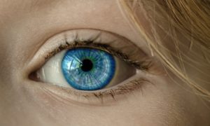
Cornea: a concave lens on the front of your eye that focuses light
Iris: the colored part, a diaphragm that controls how much light comes in (the pupil is the hole the light enters = equivalent to the aperture of a camera)
Lens: the “focuser”, uses a process called accommodation to focus near to far and make the image as sharp and clear as possible
Retina: the sensor, and sends signals to the brain to translate light into an images
The retina has 2 types of sensors:
- Rods – detect light intensity
- Cones – color differentiation
Two special areas of the retina:
- Macula – right in the middle of the retina, they place that detects the most detail (that’s why the center of your vision field is a clearer picture than the periphery)
- Fovea – the center of the macula, it contains cones (color sensors) only to aid in the translation of very fine details
Support structures
- Extra-ocular muscles – allows your eyes to move around in their holes
- There are chambers of fluid that are between each structure of the eye, and that fluid helps hold nutrients that feed those parts, and remove waste
- Choroid: the layer that holds all the blood vessels that feed the eyes
- Sclera: the whites of your eyes, an outer coating that hold everything inside
- Conjuntiva: the mucus membrane that attaches the sclera to the eyelids; produces liquid for lubrication and trapping invaders
PSA
Please don’t vigorously rub or scratch your eyes, you could hurt them!
Connect with me
Support us on Patreon
*NEW* Join the Pharmacist Answers Podcast Community on Facebook
Subscribe: iTunes, Stitcher, GooglePlay, TuneIn Radio
Music Credits: “Radio Martini” Kevin MacLeod (incompetech.com) Licensed under Creative Commons: By Attribution 3.0 http://creativecommons.org/licenses/by/3.0/