brain


Ear Anatomy [Show Notes]

Ear Basics

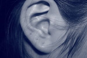
The auricle is the part of the ears you can see. It is made of cartilage (flexible tissue that doesn’t have a large blood supply).  Everything else requires a tool for the doctor to see inside.  And the doctor can only see to the ear drum.  The stuff behind the ear drum isn’t visible because of the membrane that blocks it.  The middle and inner ear are surrounded by your head bones.

Science of Sound

Sound is created when the air around us is compressed and then expands.  They move away from the source in circles (think radar or sonar or throwing a pebble in a pond).

The ear canal directs the sound waves towards the ear drum.

Sound gets translated in 2 main ways

  1. Identify the sound
  2. Identify if the sound has meaning

Inside Your Ears

The ear drum (tympanic membrane) vibrates according to the intensity of the sound and trigger the Hammer-Anvil-Stirrup cascade.

  • The ear drum vibrates the handle of the Hammer (Malus bone – yes, it’s a real bone).
  • The Hammer bangs on the Anvil (Incus bone).
  • The Anvil has a tail that is connected to the Stirrup (Stapes bone).
  • The Stirrup looks like the spurs on the back of boots.  It is connected to a membrane on the Cochlea and works like a plunger.

All of these bones are surrounded by air and the pressure is controlled by the Eustachian tube.  This is the access point for ear infections or congestion due to allergies or a cold.

The Cochlea is a bone full of fluid and lined with hairs and shaped like a spiraled sea shell.  The hairs pick up different frequencies of sound (sound wave frequency determines pitch).  If certain levels of hairs get damaged, then you will not be able to hear pitches in that range anymore.  If you unrolled the cochlea, it would be laid out low pitch to high pitch like a piano.  And these hairs are connected to the auditory nerves and turn sound signals into electrical signal to send it to your brain.

Semicircular canals of the cochlea are little bone chambers full of fluid and they control balance.  This works like a leveling bubble to help you stay upright.  If it becomes dysfunctional, then it may trigger vertigo.

The middle ear (the area behind the ear drum) is where most of the trouble happens – whether allergies causing stopped up ears, or colds leading to ear infections.

Connect with me

Support us on Patreon

*NEW* Join the Pharmacist Answers Podcast Community on Facebook

Subscribe: iTunesStitcherGooglePlayTuneIn Radio

Like the Facebook page

Music Credits:  “Radio Martini” Kevin MacLeod (incompetech.com)  Licensed under Creative Commons: By Attribution 3.0  http://creativecommons.org/licenses/by/3.0/


Male DNA in the Female Brain

*Sorry again for crying baby*

Male DNA In The News

The news reported that scientists had discovered a link between male DNA found in the brain of the women who gave birth to sons.

Microchimerism = DNA fragment of another organism that incorporates into you

This particular microchimerism involves the Y chromosome (because otherwise, you wouldn’t know it was specifically male).

Other documented microchimerism studies have reason to believe they may be beneficial – especially in a process called immuno-surveillance (when the immune system is patrolling around looking for things that don’t belong there).

The blood brain barrier is the last layer of cells between what’s in your blood and your brain cells.  DNA fragments are small and can easily pass through the BBB, especially during pregnancy when membrane permeability (the penetrable-ness) has increased all throughout the body already.

The primary resource written by the scientists that did the study of the female brains states that their findings were pretty much inconclusive – partly due to the small sample size of brains they had available.  And they couldn’t study living people.  They were mostly trying to decide if this male microchimerism had a positive or negative effect on Alzheimer’s risk.  The final conclusion – we dunno.  Another obstacle was that the complete health history of the samples they used was not known.

Other sources have stated hypotheses regarding the number of children a woman has and the risk of early onset Alzheimer’s.

This issue with reporting on studies like these is that Alzheimer’s has so many factors that may increase or decrease risk and science is pretty sure there’s NOT just one thing that will cause or prevent someone from developing this disease.

The only thing they could conclude is that microchimerisms are evolutionarily significant.

Here’s the primary journal article.

Connect with me

Support us on Patreon

*NEW* Join the Pharmacist Answers Podcast Community on Facebook

Subscribe: iTunesStitcherGooglePlayTuneIn Radio

Like the Facebook page

Music Credits:  “Radio Martini” Kevin MacLeod (incompetech.com)  Licensed under Creative Commons: By Attribution 3.0  http://creativecommons.org/licenses/by/3.0/


Now You See It [Show Notes]

Occipital Lobe Review

A brain lesion is a place in the brain that doesn’t fire when it should or fires sporadically when it shouldn’t.  Occipital lobe lesions can lead to hallucinations that range from amorphous to extremely detailed.

Field blindness: a lesion causes the occipital lobe to not translate the information from one or more spots of the visual map (your whole view).  Blind spots (round) or visual cuts (lines).

Photosensitivity seizures: seizures triggered by visual overstimulation.  Even though stereotypical in different forms of entertainment, these only accounts for about 10% of seizure triggers.  Seizures triggered by visual stimulation can range from mild to severe.

Certain types of blindness can be rooted in translation problems in the brain, rather than reception problems in the eyeball.

Lesions in the occipital-temporal-parietal junction:

  • Color agnosia: can see the colors but can’t recall the names; simplified colors (all greens appear to be the same green)
  • Movement agnosia: think weeping angels (things only move to a new position when you’re not looking at it) or moving items appear blurry
  • Agraphia: unable to communicate in writin

Connect with me

Support us on Patreon

*NEW* Join the Pharmacist Answers Podcast Community on Facebook

Subscribe: iTunesStitcherGooglePlayTuneIn Radio

Like the Facebook page

Music Credits:  “Radio Martini” Kevin MacLeod (incompetech.com)  Licensed under Creative Commons: By Attribution 3.0  http://creativecommons.org/licenses/by/3.0/


Seems like only yesterday [Show Notes]

Temporal Lobe Basics

The temporal lobe is located on each side of your head by your ears.  It helps you process auditory input and identify sounds.

There is a special area called Wernicke’s area.  It helps identify the meaning behind speech and vocal tones.  This is different from Broca’s area, which is just able to identify some sounds as being words.

This is where your long-term memory lives, like facts and knowledge (declarative).

Emotionally charged memories are also held in the temporal lobe, but they have a special connection to the amygdala (which is part of the limbic system).  These memories have a high level of detail, and usually can’t be recalled without also recalling the emotion.  They also don’t have a sense of time.

Mind Games

Memories in the temporal lobe can lead to déjà vu.

The temporal lobe allows you to see a simple or incomplete image and fill in all sorts of details, whether it be the details of the image or a long train of connected memories.

Connect with me

Support us on Patreon

*NEW* Join the Pharmacist Answers Podcast Community on Facebook

Subscribe: iTunesStitcherGooglePlayTuneIn Radio

Like the Facebook page

Music Credits:  “Radio Martini” Kevin MacLeod (incompetech.com)  Licensed under Creative Commons: By Attribution 3.0  http://creativecommons.org/licenses/by/3.0/


Where has all the grey matter gone [Show Notes]

Review

Grey matter – neuron cell bodies that create and translate messages
White matter – myelin-covered axons that transmits the messages across the brain

Conditions that affect grey matter

  1. Dementia – memory starts to fail with age (due to the death of brain cells).  Newest formed memories get lost first (Last In First Out), and it progresses until the vital functions are lost.
  2. Alzheimer’s – similar results as dementia, different cause.
  3. Bipolar – there is not a clear explanation, but the grey matter of someone who exhibits bipolar symptoms looks different on a brain scan from the grey matter of someone who doesn’t.
  4. Amnesia – can be because the cells holding the information or memories have been injured or killed due to injury, or because the wires that would send the messages for recall have been damaged.  This can be caused by head injuries.  In traumatic experiences, amnesia is a self-preserving mechanism.
  5. Lewy- body dementia – a type of dementia that manifests in Parkinson’s disease.  As a neuron cell dies, it fills up with protein and blocks message transmission.  These large clumps of protein-filled cells will show on a brain scan.  These buildups can lead to hallucinations – visual or auditory.  Also, affects memories, just like typical dementia does.
  6. Schizophrenia – stereotypical symptoms can be caused by changes in grey matter, but not the same as protein build-up.  And still a lot unknown about why.

Your brain cells do not reproduce and replenish the way other cells (like your skin) do.  We do know that the brain can create new cells, but it is a very slow process that requires very specific conditions to be present.  But the new growth of brain cells is not fast enough to slow or reverse a disease.

Connect with me

Support us on Patreon

*NEW* Join the Pharmacist Answers Podcast Community on Facebook

Subscribe: iTunesStitcherGooglePlayTuneIn Radio

Like the Facebook page

Music Credits:  “Radio Martini” Kevin MacLeod (incompetech.com)  Licensed under Creative Commons: By Attribution 3.0  http://creativecommons.org/licenses/by/3.0/


Wrinkly Brain [Show Notes]

Brain Basics

All brains are wrinkly.  Wrinkles in your brain are a good thing.  Wrinkles are biology’s way of maximizing surface area while conserving space.

The plateaus of the brain are called gyri (or a gyrus).

The smaller, sunken in wrinkles are called sulci (or a sulcus).  Sunken in sulci – that’s how I remember it.

The larger canyons of the brain are called fissures.  These are the groves that separate the hemispheres and the lobes of the brain.

Connect with me

Support us on Patreon

*NEW* Join the Pharmacist Answers Podcast Community on Facebook

Subscribe: iTunesStitcherGooglePlayTuneIn Radio

Like the Facebook page

Music Credits:  “Radio Martini” Kevin MacLeod (incompetech.com)  Licensed under Creative Commons: By Attribution 3.0  http://creativecommons.org/licenses/by/3.0/


Membranes [Show Notes]

Membranes Basics

Meninges are a triple-layer membrane that helps hold your brain in place.

The inside of your skull bones have many boney processes protruding out.  The meninges surround the brain and provide a cushioning layer around it, and anchor to these processes.  This allows the brain to be suspended inside the skull and not touch the top, sides, or bottom.

This setup is partly contributes to concussions.  When the head experiences a large enough impact, the suspended brain crashes into the bone of the skull.

Meninges encase your brain and spinal cord to protect them.

Three layers

  • Dura mater (mah-ter, not may-ter): durable and thick, contains large blood vessels.  This is the layer that anchors directly to the skull bones.  Membrane that divides hemispheres, separates a few lobes, and coats glands near the brain.
  • Arachnoid mater (yes, like spiders): wispy like spider webs – thin & transparent. Directly in contact with Dura mater, but has cellular pillars that connect it to the Pia mater.  The cerebrospinal fluid flows around these pillars. Also overs the outside of the part of the brain as a whole.
  • Pia mater – delicate, contains the capillaries that nourish the brain.  Is in direct contact with brain cells – following all the contours and wrinkles of the brain.

Subarachnoid space – hold cerebrospinal fluid (a closed fluid system that insulates and cushions the brain and spinal cord).  Doesn’t mix with blood or lymph system.

Connect with me

Support us on Patreon

*NEW* Join the Pharmacist Answers Podcast Community on Facebook

Subscribe: iTunesStitcherGooglePlayTuneIn Radio

Like the Facebook page

Music Credits:  “Radio Martini” Kevin MacLeod (incompetech.com)  Licensed under Creative Commons: By Attribution 3.0  http://creativecommons.org/licenses/by/3.0/


Caffeine and Headaches [Show Notes]

How Blood Flows

Blood vessels are like flexible pipes that run to every nook and cranny of your body.  When they dilate (or widen), they let more blood through; when they constrict (or narrow), they let less blood through.  If the blood vessels constrict and the body part at the end of that path feels deprived, it may send a pain signal to the brain.

If the blood vessels constrict or dilate quickly, your body will feel it and it may be translated as pain.

Headaches can be the result of an overall constriction of blood vessels in the head.

Your brain is full of neurons (nerve cells), and even though they translate pain, they don’t sense pain.

Caffeine Can Help

Caffeine can gently open up constricted blood vessels.

Your body is efficient, so when you sleep, certain blood vessels to certain parts of your body (like digestive tract and skeletal muscles) constrict to maximize blood flow to other places.  Once you wake up, the process by which the body re-dilates those vessels can be slow.  Exercise can make it faster.  So can caffeine.

The reason why caffeine helps us wake up: it dilates the blood vessels so more blood flows to the areas of the brain that control attention and focus and alertness.

Caffeine pill = 200 mg

Water for a headache?

If you’re dehydrated, your blood will be slightly thicker and may be harder to get to the nooks and crannies, and that can cause headache.  Dehydration can also cause low blood pressure can lead to headache.

The reason caffeine is in OTC headache pills – 1) caffeine can dilate blood vessels, and 2) it speeds up heart rate with increases how fast the other meds flow through the body.

Lucy and Ethel

Connect with me

Support us on Patreon

*NEW* Join the Pharmacist Answers Podcast Community on Facebook

Subscribe: iTunes, Stitcher, GooglePlay, TuneIn Radio

Like the Facebook page

Music Credits:  “Radio Martini” Kevin MacLeod (incompetech.com)  Licensed under Creative Commons: By Attribution 3.0  http://creativecommons.org/licenses/by/3.0/


I Think You’re Smart [Show Notes]

I think you’re smart

You’re smart, I know you are!  I want you to apply those smarts and help me out a bit.

Piles of information thrown at us about our health, some new treatment or medicine, or some new aspect of a disease.  The media is flat out MEAN about how and when it presents this information.  Usually, it’s incomplete, biased, and twisted into a scare tactic.

Anyone with a brain will know that anything in excess can be bad.

Some tips on judging the source of information

  • Are they trustworthy?  Have you even heard of them or their site?
  • What is their reputation of presenting “shock and awe” headlines just to get views, clicks, or attention?
  • Is it a celebrity or politician who wants some screen time and just wants to be the loudest voice in the room?

Critical thinking is a dying skill, please don’t let it go extinct.

Consider the Source

Science and medical information is classified based on how close to the “horse’s mouth” it is.  Scientists who do the study, write the reports and articles, and publish it in journals themselves – those are called primary sources.  To be trustworthy, they have to state any limitations their study had, confounding factors, and any biases (aka money paid by someone who cares, like drug companies or lobbying groups).  Then there may be a group of scientists or statisticians that take several primary resources that studied the same thing and compare all their outcomes and come up with an overarching conclusion – those are called secondary sources.  Articles that reach the public and may have citations of using primary or secondary sources for some of the information presented are called tertiary sources.  They are so far removed from the primary source that they CANNOT be used for academic research!

Again, critical thinking skills are vital for our success in society.  Don’t follow the fads!  Don’t allow yourself to be duped by people who line their pockets with money from people who are ignorant about the subject they’re screaming.

Connect with me

Support us on Patreon

*NEW* Join the Pharmacist Answers Podcast Community on Facebook

Subscribe: iTunes, Stitcher, GooglePlay, TuneIn Radio

Like the Facebook page

Music Credits:  “Radio Martini” Kevin MacLeod (incompetech.com)  Licensed under Creative Commons: By Attribution 3.0  http://creativecommons.org/licenses/by/3.0/


Location of Important Organs

Ever have a twinge and wonder what organs or body parts are in that area?  When the next step is to run a Google symptom search, you will find yourself faced with a potentially deadly diagnosis in approximately 3.2 seconds!
To avoid the unnecessary stress and anxiety of appointments with Dr. Google, help yourself make wise health decisions by knowing the approximate location of your organs.

Important Organs

Brain:  It’s in your head (obviously).  Interesting tidbit: your main is made of nerves but they are not sensory neurons, so your brain doesn’t feel.  The other things around your brain feel, like membranes and connective tissue inside and outside the skull.
Thyroid:  It is on the front of your neck right above the middle dip between your collar bones.
Heart:  Ball of your fist and put it right in the middle of your chest (yes ladies, right between your boobs).
Lungs:  They actually start way up high right below your collarbones and extend down and stop right about your last complete rib.
Stomach:  It starts in the middle, right under your sternum (or breastbone) and curves to the left. Interesting tidbit: laying on your left side causes gastric emptying, which is when the stomach dumps its contents into the top of the small intestines.
Liver:  It is shaped like a triangle on your right side, starts under your ribs. The long side of the triangle crosses over what you might call “the pit of your stomach”, the short side extends down about even with your belly button.
Gallbladder:  It’s located under the liver, a little to the right of “the pit of your stomach”.
Small intestine:  It snakes its way left and right across your abdominal region
Kidneys:  They are towards your back, under the bottom few ribs.
Colon:   It is right above your tailbone.  Interesting tidbit: this is why constipation can cause back pain.
Appendix:  It is on your right side, above your hip bone.
Bladder:  It is right above the pubic bone (the middle bone that connects the two sides of the pelvis). I mean, we all know what it feels like to have to pee, right?
So this is a list of the major things that you can’t see but you might be able to feel if something isn’t quite right.