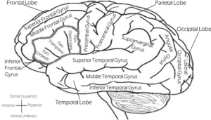
Brain


See it in my head [Show Notes]

Parietal Lobe

It sits in the top of your head

  • Responsible for translating: touch, temperature, and pain.
    • So does it tickle? Or hurt?  How does it hurt – throbbing, stabbing, sharp, dull?
      The way your body translates temperature is based on the relative temperature to the body part that is being touched.
  • Proprioception – ability to know where you are in space relative to the other things around you
  • That awkward moment  when you think the toilet is higher or closer that it really is and you almost fall.
  • Hand-eye coordination – being able to see something that is moving and make your body to interact with it.
  • Two-point discrimination – the body’s ability to tell if it’s being touched by one thing or two separate things.  Different parts of your body have different levels of sensitivity.  Large body parts have a larger distance than small body parts (fingers and tongue have the closest discrimination distance due to the number of sensors).
  • Graphesthesia = writing feelings = being able to correctly translate letters or shapes drawn on your body without you looking.

When someone says “I can see it in my head” – it’s the area that can recall visual memories.  Remembering the last place you saw your keys or visualizing driving directions based on the landmarks you pass (which is the only way we give directions in the South).  So, when someone closes their eyes to try and remember something, they’re trying to deactivate their eyes and activate the parietal lobe.

Being able to identify a 3D object with your hands by touch only, and without your eyes.  This is also how Braille works, the dot patterns created to represent letters.

Good info here and here about Braille activates the brain.


On the tip of my brain [Show Notes]

Recap

Frontal Lobe

  • Motor cortex – voluntary muscle movements, including the muscles that control speech
  • Language translation
  • Prefrontal cortex – personality, judgment

Dopamine

The main chemical, or neurotransmitter, that functions in the frontal lobe is dopamine.

Reward System and Addiction

  • Dopamine is part of your brain’s reward system.
    • So think about when you get a Facebook notification…. dopamine is released in your brain, and your brain really likes how dopamine makes it feel.  Feels good!  So you’re brain will help you pay more attention to the things that will get you more dopamine (that’s why a 5-second Facebook glance can turn into 30 minutes).  This also means that dopamine is involved in your attention span.
    • Some newer studies are looking at dopamine’s effect on addiction.
    • The problem is that it requires increasing levels of “excitement” for your brain to receive the same level of dopamine as the very first time.  This is why people with devastating addictions end up on a downward spiral of ruin.

Memories

  • Dopamine is also involved in short-term memory, especially in complex or cascading tasks (where you have to remember a thing from Step A to complete Step B) in your prefrontal cortex.
    • Diseases that take away short-term memory:  Dementia (general or Parkinson’s-related), Alzheimers.
    • Dopamine is being studied in how it related to dementia and Alzheimer’s.  It’s effects are already known in Parkinson’s disease.
    • To form memories, your brain has to access the same information over and over again (like a smooth, speedy highway).  A road only traveled once, is not easy to travel again, especially if there’s a long period of time between trips down that road.  So in diseases that involve brain cell death, there becomes less and less routes to take to the same memory.  Thus, the older memories are the last to go because they have the most access routes.

Planing

  • Dopamine is responsible for your planning and motivation mechanisms.  If I make a plan and carry out the plan, the reward of dopamine is the outcome.

A New Discovery

They’ve discovered a genetic component that affects the shape of the dopamine receptors.

scanned-image

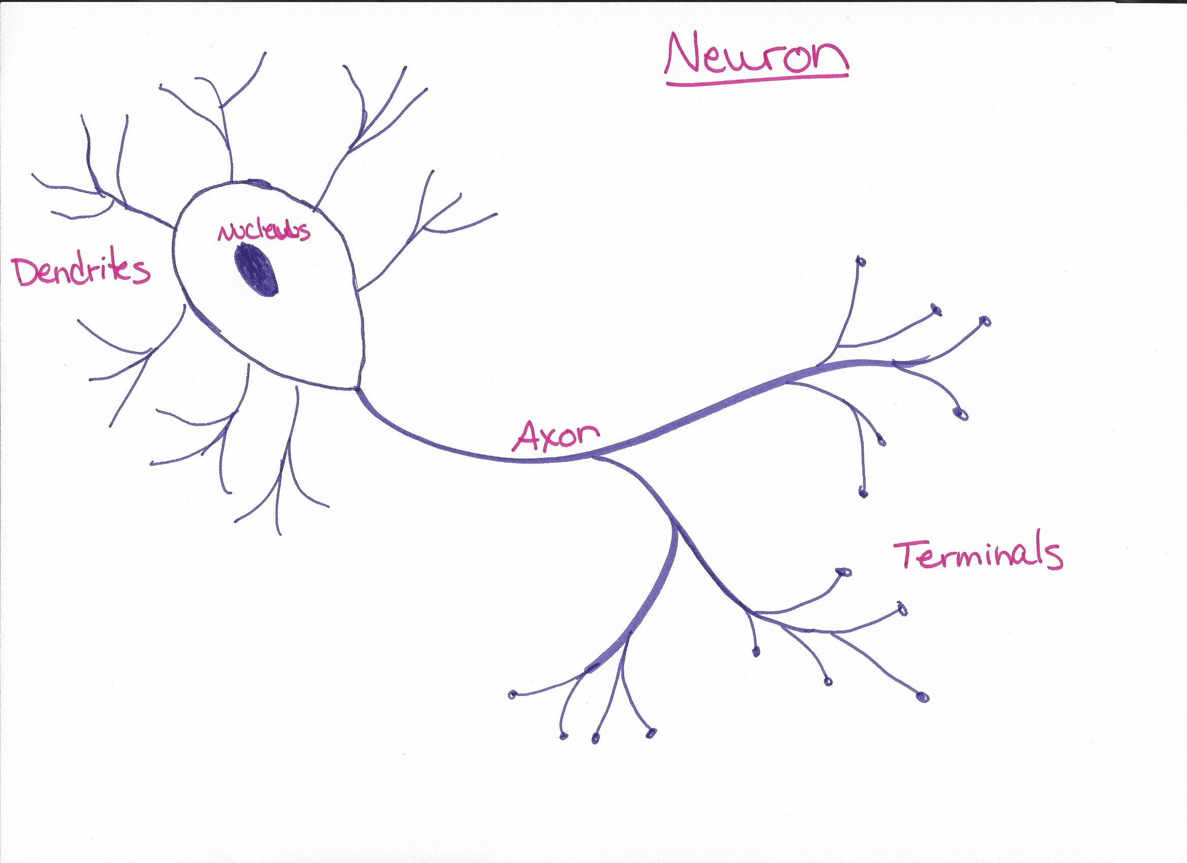
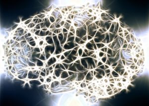
These cells in your brain don’t actually touch each other.  The terminals spit out dopamine, and it floats in the space and hopefully comes in contact with the next cell’s dendrites.  The dendrites have dopamine-shaped keyholes, and the dopamine should fit in the keyholes perfectly.  But they have discovered that a genetic component affects the keyhole shapes, and this make be a root cause for schizophrenia and attention deficits.

If you think about it, the classic symptoms of schizophrenia – paranoia, anxiety, hallucinations, split personalities – most things affected in your prefrontal cortex.  So if the dopamine receptors are “broken” in this area of your frontal lobe, you can see how there could be a dysfunction.  And this is produced at the genetic level.  Science is still learning about this…

Stroke

Strokes or brain injury in this area of your brain can affect personality. These parts of your brain don’t grow back! Some issues related to the speech motor area of the brain (Broca’s area).  Stuttering (clinically diagnosed) is a misfire in the motor planning part of speech.  Aphasia (loss of words) – part of you brain knows the word but you can’t seem to get it out of your mouth.  Strokes in this area can cause some strange effects in the loss of words.

Seizures

There is also a type of epilepsy (seizures).  Seizures are a misfire or a short in the electrical signals of the brain.  Seizures in the frontal lobe can possibly affect memory (epilepsy-related amnesia).  Must be diagnosed by a neurologist.

Story Time

Back in the day, there was a guy named Pheneas Gage who worked on the railroad.  An accident involving dynamite and a railroad spike, led to a major head injury and an altered personality!

We rub our forehead when we’re trying to remember something because that’s where our short-term memory is.

#RealTalk

Cynthia doesn’t have that many Facebook friends!

Connect with me

Support us on Patreon

*NEW* Join the Pharmacist Answers Podcast Community on Facebook

Subscribe: iTunesStitcherGooglePlayTuneIn Radio

Like the Facebook page

Music Credits:  “Radio Martini” Kevin MacLeod (incompetech.com)  Licensed under Creative Commons: By Attribution 3.0  http://creativecommons.org/licenses/by/3.0/


Makes You Who You Are [Show Notes]

**Many apologies for the screaming baby***

Basics

Frontal Lobe – in the front of the brain (behind your forehead)

This is the area that contains your personality.  A stroke or brain injury or damage can alter someone’s personality drastically.

We take personality tests, but they are too basic to take such a complex part of you and put it in a quadrant or on a spectrum.

This area of the brain is also well-connected to the limbic system (emotional center), and if those connections are broken through stroke or injury, then that causes personality changes too.

Frontal Lobe

  • Motor cortex: voluntary muscle movements = how you choose to move your body.
  • Prefrontal cortex: personality, complex cognitive behavior, decision-making, social behavior, judgment (not existential judgement – but simple things like opposites) – if this gets damaged in adults (so less chance to relearn things as kids would), they don’t sense the dread of the consequences of doing “bad” behaviors, thus they can live lifestyles that include sex, drugs, and crime.
  • Broca’s area (confirmed: he was French):  speech, language production, translation (not just audible language, but any type of symbol or gesture that would have meaning)
    • stuttering and aphasia originate here

* This area of the brain doesn’t reach full development until almost 30 years old —> Insert rant here!!

Story Time

During the Heroic Era of medicine (not a well-named era) – they invented the Lobotomy – mainly trying to find a treatment for mental illnesses.  Society was ok with doctors experimenting on criminals in prison and patients who were put in asylums by their families. It did cause changes in the people, thus they claimed the “cured” them.

Connect with me

Support us on Patreon

*NEW* Join the Pharmacist Answers Podcast Community on Facebook

Subscribe: iTunesStitcherGooglePlayTuneIn Radio

Like the Facebook page

Music Credits:  “Radio Martini” Kevin MacLeod (incompetech.com)  Licensed under Creative Commons: By Attribution 3.0  http://creativecommons.org/licenses/by/3.0/


White Matter Matters [Show Notes]

Review

Disease like Alzheimer’s and dementia are grey matter issues – the creation and translation of messages are interrupted or dysfunctional.  White matter is like the power cords that are responsible for sending the signals.

White matter diseases

  • Hypomylenation – cells are created with a low amount of myelin; premature, chromosome-linked defects
    • Cell Biology review:  animal cells have a membrane and a nucleus that holds all the DNA, and cytoplasm, and then all the organelles that have different jobs – just like a self-contained factory.  Some systems can be dysfunctional and the cell still live and replicate.
  • Dysmylenation – neurodystrophy (a huge list based on what’s broken)
    • Lysosomes – stores enzymes for breakdown
    • Perioxosomes – stores enzymes for energy metabolism
    • Mitochondrial – dysfunction of energy usage
    • Amino acid metabolism dysfunction
  • Demylenation
    • Inflammatory: Multiple Sclerosis = autoimmune disease.  The brain wants the body to do something but the message doesn’t make it to the body, so the body doesn’t move or has very jerky, irregular movements.  Tests for antibodies can identify MS.  Available treatment is mostly immune suppressants.
    • Huntington’s is a genetic disease that presents in the same way.  It has a very sad prognosis and presents in females starting between the ages of 30 and 50.  Thus they have already planned a life and possibly had kids who now may have the same disease.  Genetic testing can identify Huntington’s.
    • Viral – PML (Progressive Multifocal Leukoencephalopathy); J-virus a typical virus that may mutate and go dormant in the brain.  If the immune system is lowered drastically – due to suppression or immunodeficiency diseases, this virus will wake up and attack the myelin of the neurons.
    • Acquired metabolism demyleniation (being exposed to chemicals) – “Chasing the Dragon” – refers to a technique used to keep melted pills from burning in a container (usually a spoon) while it’s being heated by a flame from underneath and the vapors are inhaled.  Drug of choice: heroin.  **DON’T DO DRUGS**
    • Hypoxic ischemic – loss of oxygen.  Examples:  asphyxiation, drowning, ischemic stroke.
    • Mechanical – compression due to injury or swelling

Call Back

Migraines are not a white matter issue, they are a brain chemistry issue.  Learn more on the Headache episode

Connect with me

Support us on Patreon

*NEW* Join the Pharmacist Answers Podcast Community on Facebook

Subscribe: iTunesStitcherGooglePlayTuneIn Radio

Like the Facebook page

Music Credits:  “Radio Martini” Kevin MacLeod (incompetech.com)  Licensed under Creative Commons: By Attribution 3.0  http://creativecommons.org/licenses/by/3.0/


Where has all the grey matter gone [Show Notes]

Review

Grey matter – neuron cell bodies that create and translate messages
White matter – myelin-covered axons that transmits the messages across the brain

Conditions that affect grey matter

  1. Dementia – memory starts to fail with age (due to the death of brain cells).  Newest formed memories get lost first (Last In First Out), and it progresses until the vital functions are lost.
  2. Alzheimer’s – similar results as dementia, different cause.
  3. Bipolar – there is not a clear explanation, but the grey matter of someone who exhibits bipolar symptoms looks different on a brain scan from the grey matter of someone who doesn’t.
  4. Amnesia – can be because the cells holding the information or memories have been injured or killed due to injury, or because the wires that would send the messages for recall have been damaged.  This can be caused by head injuries.  In traumatic experiences, amnesia is a self-preserving mechanism.
  5. Lewy- body dementia – a type of dementia that manifests in Parkinson’s disease.  As a neuron cell dies, it fills up with protein and blocks message transmission.  These large clumps of protein-filled cells will show on a brain scan.  These buildups can lead to hallucinations – visual or auditory.  Also, affects memories, just like typical dementia does.
  6. Schizophrenia – stereotypical symptoms can be caused by changes in grey matter, but not the same as protein build-up.  And still a lot unknown about why.

Your brain cells do not reproduce and replenish the way other cells (like your skin) do.  We do know that the brain can create new cells, but it is a very slow process that requires very specific conditions to be present.  But the new growth of brain cells is not fast enough to slow or reverse a disease.

Connect with me

Support us on Patreon

*NEW* Join the Pharmacist Answers Podcast Community on Facebook

Subscribe: iTunesStitcherGooglePlayTuneIn Radio

Like the Facebook page

Music Credits:  “Radio Martini” Kevin MacLeod (incompetech.com)  Licensed under Creative Commons: By Attribution 3.0  http://creativecommons.org/licenses/by/3.0/


Wrinkly Brain [Show Notes]

Brain Basics

All brains are wrinkly.  Wrinkles in your brain are a good thing.  Wrinkles are biology’s way of maximizing surface area while conserving space.

The plateaus of the brain are called gyri (or a gyrus).

The smaller, sunken in wrinkles are called sulci (or a sulcus).  Sunken in sulci – that’s how I remember it.

The larger canyons of the brain are called fissures.  These are the groves that separate the hemispheres and the lobes of the brain.

Connect with me

Support us on Patreon

*NEW* Join the Pharmacist Answers Podcast Community on Facebook

Subscribe: iTunesStitcherGooglePlayTuneIn Radio

Like the Facebook page

Music Credits:  “Radio Martini” Kevin MacLeod (incompetech.com)  Licensed under Creative Commons: By Attribution 3.0  http://creativecommons.org/licenses/by/3.0/


White matter vs Grey Matter [Show Notes]

Basic Brain Biology

Your brain is made of cells.  Those cells are called neurons.  Neurons transmit signals in the form of electricity (aka .positive and negative charges).  One end of a neuron will build a signal or charge, and once it reaches a certain threshold, then a signal is send down the axons.

Most of the cells in your body touch and transmit signals and pass chemicals through their membranes.  Neurons do not touch.  The terminals of one will get really really close to the dendrites of another.

They’re really good at the telephone game – mostly because the body tries to minimize the number of neurons involved in passing a signal.

Axons are coated in myelin.  Myelin insulates the axon that helps the signal being sent travel faster, and prevents it from getting lost to something else touching it.  You want the signal to have to same strength when it reaches its destination as it did when it left its source.

Parts of a neuron

Dendrites: receives signals from previous neuron
Cell body: contains the nucleus and creates and translates signals
Axon: the “wire” that transmits signals
Terminals: sends signals to the next neuron

a hand-drawn sketch of a basic neuron with labeled parts. This makes up white matter and grey matter in the brain.

Grey matter – cell bodies, dendrites, and terminals
White matter – axons wrapped in myelin

Grey matter – information storage and translation
White matter – information transmission

Brain: grey matter is on the outside, white matter is on the inside
Spinal cord:  grey matter is on the inside, white matter is on the outside.

PS.  Grey?  Gray?  IDK!!!

Connect with me

Support us on Patreon

*NEW* Join the Pharmacist Answers Podcast Community on Facebook

Subscribe: iTunesStitcherGooglePlayTuneIn Radio

Like the Facebook page

Music Credits:  “Radio Martini” Kevin MacLeod (incompetech.com)  Licensed under Creative Commons: By Attribution 3.0  http://creativecommons.org/licenses/by/3.0/


Meningitis [Show Notes]

Basics

Meningitis = inflammation of meninges

Generalized symptoms:  fever, headache, stiff neck, light sensitivity, confusion, lethargy, 

4 types of infectious meningitis

  1. Bacterial – we have a vaccine for that! 3-7 days; spreads in close communities (like college dorms).  Spreads through prolonged contact.  Confirmed through spinal tap and cerebrospinal fluid culture
  2. Viral – most common type; can be caused by lots of viruses: enterovirus, mumps, measles, flu, west nile.  7-10 days.  This is why I believe in vaccines!
  3. Fungal – not contagious; most common in patient with suppressed immune systems or secondary from surgery.
  4. Parasitic – Rare yet fatal.  Amoeba enters through nose from warm contaminated fresh water sources or pool, even hot springs (warm up to 115* F).  Only 31 cases in the 10 years between 2002 and 2012.  Destroys brain tissue ~ 5 days. 

Non-infectious causes:  Surgery, injury, lupus, cancer

The most important thing is that all of this be monitored by a doctor – ALWAYS!!

2 quick tests for meningitis

Kernig’s sign 

Brudzinski’s sign

Connect with me

Support us on Patreon

*NEW* Join the Pharmacist Answers Podcast Community on Facebook

Subscribe: iTunesStitcherGooglePlayTuneIn Radio

Like the Facebook page

Music Credits:  “Radio Martini” Kevin MacLeod (incompetech.com)  Licensed under Creative Commons: By Attribution 3.0  http://creativecommons.org/licenses/by/3.0/


Brain Bleeds [Show Notes]

Review

Your brain is held inside your skull by a tri-layer membrane called the meninges.  These membranes and all the other structures in your brain are nourished by blood vessels, and different circumstances will make these vessels at risk of rupturing.

*ALL BRAIN BLEEDS REQUIRE MEDICAL ATTENTION!* 

Brain bleeds are classified based on the membrane they are closest to.  They can be caused by physiological malformations, stroke or aneurism from age or disease, or trauma.

From the outside in

  • Extracranial bleed: (extra = external; cranial = cranium = skull bones), between your skin and your skull.  Doesn’t affect your brain, there is more room for it to stretch.
  • Intracranial bleed: (intra = internal); bleeds inside the skull increase the intracranial pressure and requires medical intervention.  Main goal is to reduce intracranial pressure so brain cells aren’t pressed on and damaged.
    • Epidural (yep, that place they put the anesthesia for women having babies, except it’s in the spinal cord): Epi = above, Dural = Dura mater, that topmost, durable layer of the meninges.  Between the skull and the dura mater.
    • Subdural: Sub = under; blood leaks in between the dura mater and the arachnoid mater, which are normally in close contact, so the separation causes pain.
    • Subarachnoid: under the arachnoid mater. Normally, under the arachnoid layer is the subarachnoid space which contain cerebral spinal fluid (CSF).  People who have had a subarachnoid bleed and survived, describe hearing a “thunderclap”.  Officially called a “thunderclap headache”.  It’s like they can hear the blood vessel pop and experience extreme pain all over their head all at once.  Described as “the worst headache of my entire life”.  Because the pia mater under the subarachnoid space lays directly on top of the brain cells and follows all the grooves and wrinkles of the brain, this type of bleed will require emergent attention and possible surgery.
  • Intracerebral: cerebral = cerebrum, the main part of your brain
  • Intraparenchymal: Parenchyma = organ tissue, means it’s right up against the brain cells
  • Intraventricular: Ventricles = pockets inside the brain that make, hold, and reabsorb CSF.  The deepest part of the brain.

*ALL BRAIN BLEEDS REQUIRE MEDICAL ATTENTION!* 

Connect with me

Support us on Patreon

*NEW* Join the Pharmacist Answers Podcast Community on Facebook

Subscribe: iTunesStitcherGooglePlayTuneIn Radio

Like the Facebook page

Music Credits:  “Radio Martini” Kevin MacLeod (incompetech.com)  Licensed under Creative Commons: By Attribution 3.0  http://creativecommons.org/licenses/by/3.0/


Membranes [Show Notes]

Membranes Basics

Meninges are a triple-layer membrane that helps hold your brain in place.

The inside of your skull bones have many boney processes protruding out.  The meninges surround the brain and provide a cushioning layer around it, and anchor to these processes.  This allows the brain to be suspended inside the skull and not touch the top, sides, or bottom.

This setup is partly contributes to concussions.  When the head experiences a large enough impact, the suspended brain crashes into the bone of the skull.

Meninges encase your brain and spinal cord to protect them.

Three layers

  • Dura mater (mah-ter, not may-ter): durable and thick, contains large blood vessels.  This is the layer that anchors directly to the skull bones.  Membrane that divides hemispheres, separates a few lobes, and coats glands near the brain.
  • Arachnoid mater (yes, like spiders): wispy like spider webs – thin & transparent. Directly in contact with Dura mater, but has cellular pillars that connect it to the Pia mater.  The cerebrospinal fluid flows around these pillars. Also overs the outside of the part of the brain as a whole.
  • Pia mater – delicate, contains the capillaries that nourish the brain.  Is in direct contact with brain cells – following all the contours and wrinkles of the brain.

Subarachnoid space – hold cerebrospinal fluid (a closed fluid system that insulates and cushions the brain and spinal cord).  Doesn’t mix with blood or lymph system.

Connect with me

Support us on Patreon

*NEW* Join the Pharmacist Answers Podcast Community on Facebook

Subscribe: iTunesStitcherGooglePlayTuneIn Radio

Like the Facebook page

Music Credits:  “Radio Martini” Kevin MacLeod (incompetech.com)  Licensed under Creative Commons: By Attribution 3.0  http://creativecommons.org/licenses/by/3.0/