Monthly Archives: January 2017


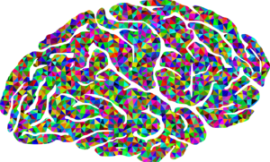
An Accordion in Your Brain

Cerebellum Basics

Your cerebellum is a separate part of your brain that sits under the occipital lobe.  It is responsible for unconscious motor functions, and is organized differently than the cerebrum.  It is packed tightly together in neat folds like an accordion.  And it has 3 lobes:

  • Anterior (in the front) – it keeps the body visually “centered” and on balance, as well as moving the head or body to keep the eyes level with the horizon.  Alcoholism can cause damage to this area that results in a person being sober but still walking “drunk”.
  • Follcular-nodular (in the middle with nodules on it) – responsible for eye movement in response to motion.  It is responsible for correcting balance based on signals from the body rather than the eyes. This is how you know you’re falling over when you have your eyes closed.  Also responsible for muscle tone (aka the passive contraction or “readiness” of a relaxed muscle).
  • Posterior (in the back) – responsible for fine motor coordination and it turns off signals for involuntary movements.

Purkinje cells are the main type of neuron in the cerebellum – SO BEAUTIFUL!!

Connect with me

Support us on Patreon

*NEW* Join the Pharmacist Answers Podcast Community on Facebook

Subscribe: iTunesStitcherGooglePlayTuneIn Radio

Like the Facebook page

Music Credits:  “Radio Martini” Kevin MacLeod (incompetech.com)  Licensed under Creative Commons: By Attribution 3.0  http://creativecommons.org/licenses/by/3.0/


Now You See It [Show Notes]

Occipital Lobe Review

A brain lesion is a place in the brain that doesn’t fire when it should or fires sporadically when it shouldn’t.  Occipital lobe lesions can lead to hallucinations that range from amorphous to extremely detailed.

Field blindness: a lesion causes the occipital lobe to not translate the information from one or more spots of the visual map (your whole view).  Blind spots (round) or visual cuts (lines).

Photosensitivity seizures: seizures triggered by visual overstimulation.  Even though stereotypical in different forms of entertainment, these only accounts for about 10% of seizure triggers.  Seizures triggered by visual stimulation can range from mild to severe.

Certain types of blindness can be rooted in translation problems in the brain, rather than reception problems in the eyeball.

Lesions in the occipital-temporal-parietal junction:

  • Color agnosia: can see the colors but can’t recall the names; simplified colors (all greens appear to be the same green)
  • Movement agnosia: think weeping angels (things only move to a new position when you’re not looking at it) or moving items appear blurry
  • Agraphia: unable to communicate in writin

Connect with me

Support us on Patreon

*NEW* Join the Pharmacist Answers Podcast Community on Facebook

Subscribe: iTunesStitcherGooglePlayTuneIn Radio

Like the Facebook page

Music Credits:  “Radio Martini” Kevin MacLeod (incompetech.com)  Licensed under Creative Commons: By Attribution 3.0  http://creativecommons.org/licenses/by/3.0/


Eyes In The Back of Your Head [Show Notes]

Occipital Lobe Basics

The occipital lobe sits in the back of your head, it directly connects to your eyes.

2 Streams of messages that your eyes send to your occipital lobe.

  1. Ventral stream – translates “what”
  2. Dorsal stream – translates “where” and “how”

It sends translated information to the necessary part of the brain to respond or react to what you saw.  This is how hand-eye coordination works (not just for athletes).  And, since so much of the information we receive is visual means that the occipital lobe doesn’t do much else.

**The following is complete speculation based on my experiences as a Mom.**

Mom’s get accused of having eyes in the back of their head – but my guess is that mom’s gain a keener sense of spacial awareness regarding the things that are happening around you.  Also, mom’s hearing become much more attuned to specific sounds (aka knowing their baby’s voice from other baby voices) to the point of knowing the difference between the sound of crayons coloring on paper versus crayons coloring on a wall!

If it hasn’t been obvious, let me just say that no part of the brain acts and reacts all by itself.  Many of the complex activities we complete as humans involve many areas of the brain simultaneously or sequentially.

Connect with me

Support us on Patreon

*NEW* Join the Pharmacist Answers Podcast Community on Facebook

Subscribe: iTunesStitcherGooglePlayTuneIn Radio

Like the Facebook page

Music Credits:  “Radio Martini” Kevin MacLeod (incompetech.com)  Licensed under Creative Commons: By Attribution 3.0  http://creativecommons.org/licenses/by/3.0/


Art & Vitamins [Show Notes]

Temporal Lobe Problems

Temporal lobe lesions can lead to dyslexia.

Receptive aphasia: can’t receive or translate speech meaning

Word deafness: words are only noise

Temporal lobe lesions can also lead to deafness.  The ears are fine, but the wires that translate input as sound are damaged. (Possibly what happened to Helen Keller).

Callbacks

Meningitis

Brain Bleeds

Big Words

Prospoagnosia = facial blindness

Chuck Close

Clinical apathy: you forget how to feel

Anterograde amnesia: can’t make new memories
Retrograde amnesia: can’t recall past memories
Situational amnesia: self-preservation from trauma

Wernicke-Korsakoff Syndrome

  • Vitamin B-1 (Thiamine) deficiency
    • alcoholics
    • careless vegetarian/vegan diets
  • Lose ability to walk, talk, and remember

Connect with me

Support us on Patreon

*NEW* Join the Pharmacist Answers Podcast Community on Facebook

Subscribe: iTunesStitcherGooglePlayTuneIn Radio

Like the Facebook page

Music Credits:  “Radio Martini” Kevin MacLeod (incompetech.com)  Licensed under Creative Commons: By Attribution 3.0  http://creativecommons.org/licenses/by/3.0/


Seems like only yesterday [Show Notes]

Temporal Lobe Basics

The temporal lobe is located on each side of your head by your ears.  It helps you process auditory input and identify sounds.

There is a special area called Wernicke’s area.  It helps identify the meaning behind speech and vocal tones.  This is different from Broca’s area, which is just able to identify some sounds as being words.

This is where your long-term memory lives, like facts and knowledge (declarative).

Emotionally charged memories are also held in the temporal lobe, but they have a special connection to the amygdala (which is part of the limbic system).  These memories have a high level of detail, and usually can’t be recalled without also recalling the emotion.  They also don’t have a sense of time.

Mind Games

Memories in the temporal lobe can lead to déjà vu.

The temporal lobe allows you to see a simple or incomplete image and fill in all sorts of details, whether it be the details of the image or a long train of connected memories.

Connect with me

Support us on Patreon

*NEW* Join the Pharmacist Answers Podcast Community on Facebook

Subscribe: iTunesStitcherGooglePlayTuneIn Radio

Like the Facebook page

Music Credits:  “Radio Martini” Kevin MacLeod (incompetech.com)  Licensed under Creative Commons: By Attribution 3.0  http://creativecommons.org/licenses/by/3.0/